Pollen Microscopy – taking a closer look at spring forage
This weekend I have been using pollen microscopy to identify the different spring forage being brought into the apiary. Foraging bees were bringing in several different colours of Pollen. These ranged from bright orange through bright yellow, to light yellow. With so much forage available, here in rural Northumberland, I was keen to identify what the foragers were bringing back to the hive.
What approach did I use for Pollen Identification?
The approach I used for Pollen Identification involved:
- Looking to see what was in flower in the surrounding area in significant quantity
- Trying to see which flowers the bees were visiting
- Taking samples of the pollen from these plants
- Recording the colour of the pollen loads arriving on the landing boards
- Making up pollen slides and examining them under the microscope
- Confirming the colour of the pollen load with Rex Sawyers Pollen Identification
The outside photographs in this blog were taken with a Nikon D3200, fitted with a Tamron 18-275 mm lens and a Sigma 105mm macro lens for the close-up shots. The microscope photos were taken with an apex minigrab camera, fitted into the eyepiece of my Apex Researcher compound microscope.
Identifying Dandelion using Pollen Microscopy
There is a large amount of dandelion in flower. It is along all of the hedgerows, in open grassland and in the apiary. It is hated by most gardeners, but the bees love it.
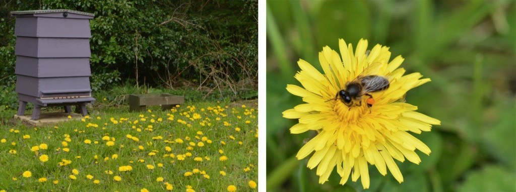
Watching the flowers in the apiary, foragers could be seen with bright orange pollen in their baskets. The same distinctive colour could also be seen on the landing board of the hive. The short journey time must have made it very appealing this weekend.
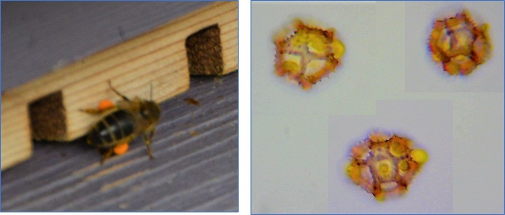
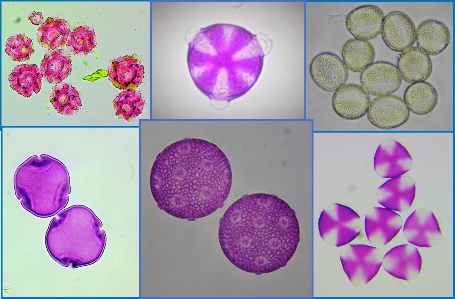
Dandelion pollen is very distinctive. It’s an easy one to remember for anyone studying for their BBKA Microscopy exam. It usually has a lot of the yellow oily substance on and around the pollen grains. The pollen varies in size around 30 um. It is very roughly round, but it’s surface is made up of very distinctive, jagged, ridges. These ridges have spines all along them.
Identifying Oil Seed Rape using Pollen Microscopy
The oil seed rape is in flower and the foragers were making a bee-line for it. It is a favourite of the honey bee but surprisingly was not the only pollen arriving on the landing board.
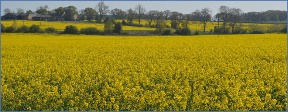
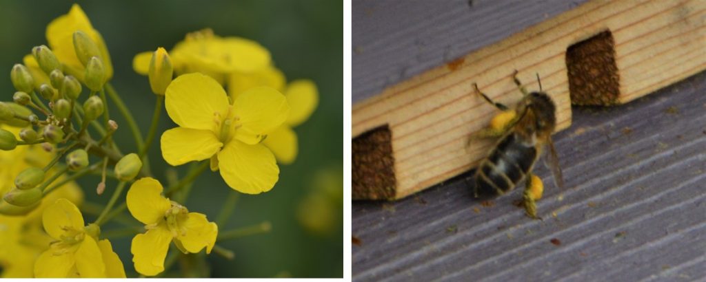
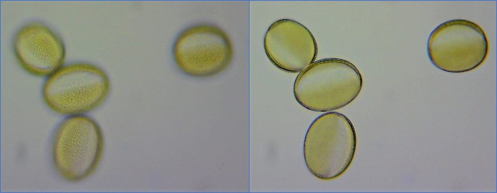
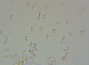
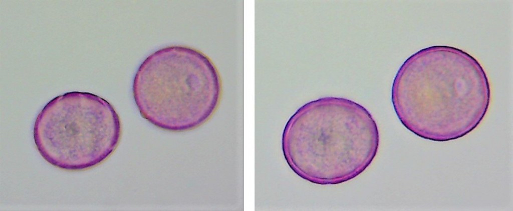
The Oil Seed Rape pollen measured 38um under the microscope and is oblong in shape. The pores are not prominent. The two pictures below show the photos taken through the eyepiece. The one on the left shows the netted and beaded surface of the pollen grain. The one on the right shows the section at the equator and the medium thick exine (shell).
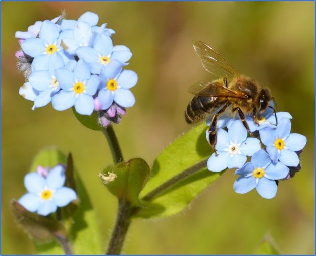
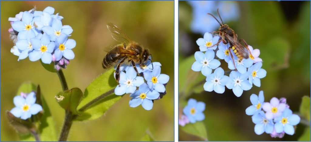
Identifying Forget Me Not using pollen microscopy
Forgot Me Not is currently in full flower. It is hard to imagine that it’s tiny little flowers yield enough to interest bees. So, it was interesting to watch a patch of it to see how much interest it was generating across different species, including honey bees.
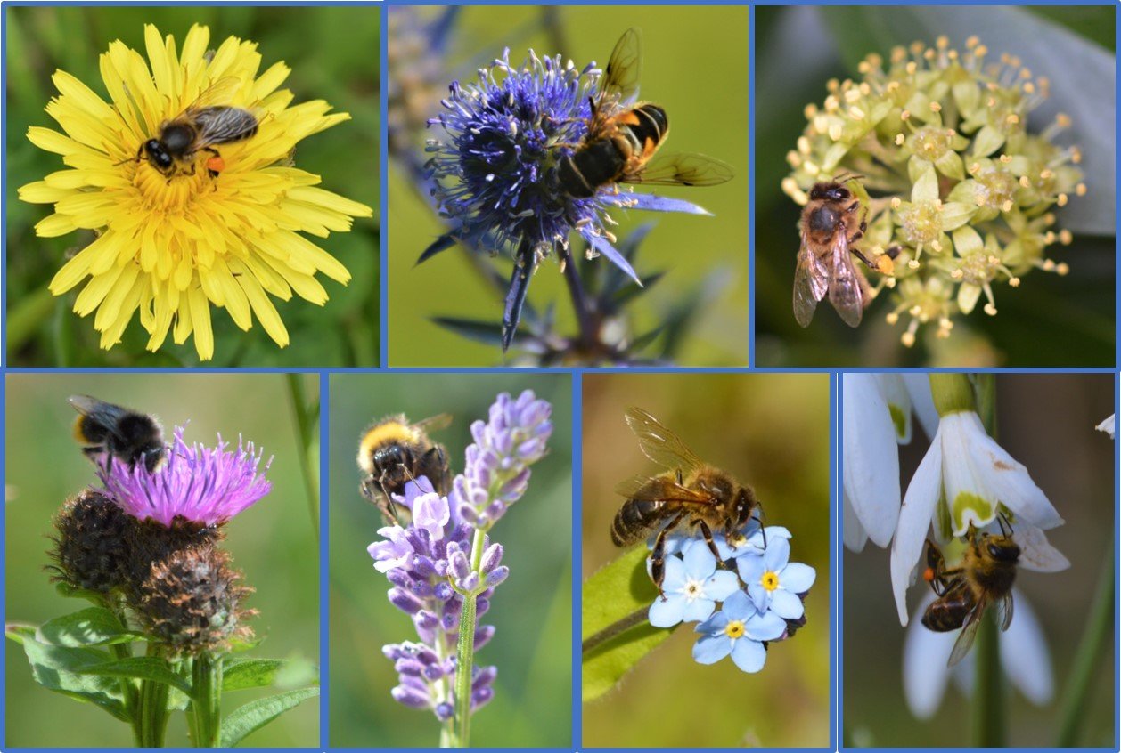
The picture on the left shows a honey bee foraging for nectar, with its proboscis extended into the narrow nectary of the forget me not. The right-hand side photo shows a species of solitary bee, also working the Forget Me Not. From Steven Falk’s Field Guide to British Bees, I think this is a Gooden’s nomad bee.
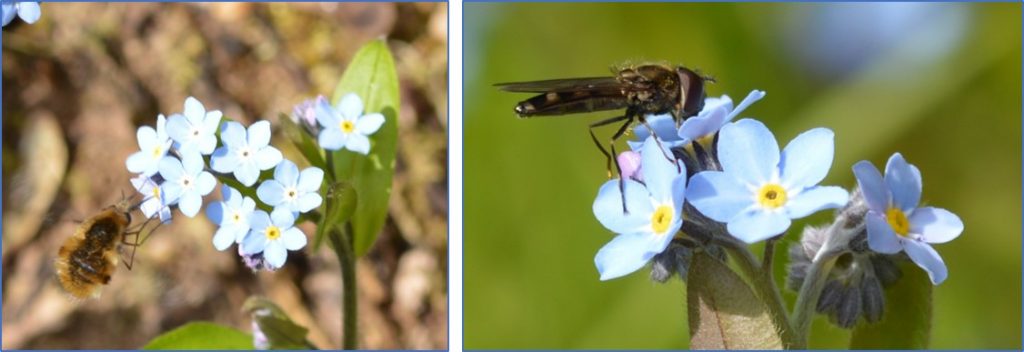
There were also species of fly working the Forget Me Not, including this Bee-Fly on the left and Hover Fly on the right, below.
Forget Me Not pollen is tiny, at about 6um. It is a very distinctive dumbbell shape. Rex Sawyers pollen identification guide shows that Forget Me Not pollen is a yellow colour. There were different shades of yellow pollen going into the hive and the bees were working this flower, but it looked like they were taking nectar and none of the honey bees had pollen in their baskets.
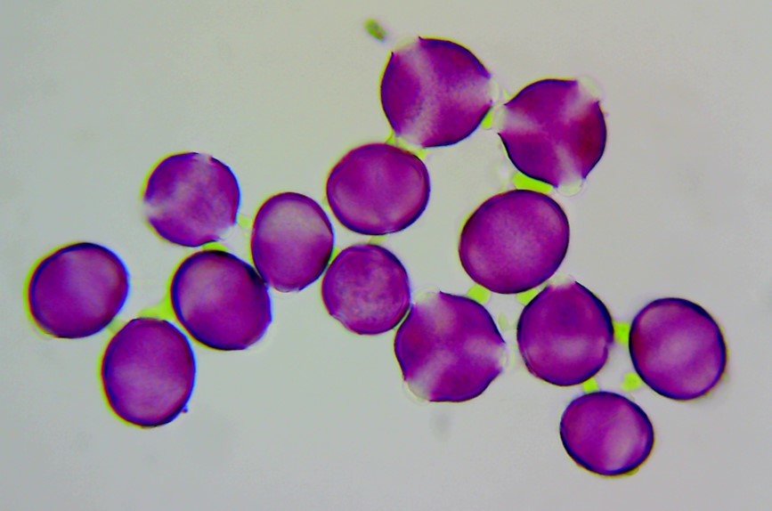
Identifying Black Thorn using Pollen Microscopy
The black thorn in the woods and hedgerows is in flower in the area, in abundance.
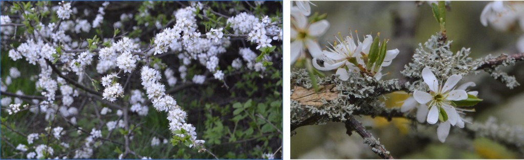
Black thorn pollen is round/oblong in shape and is about 48um in size. The photo on the left shows the thin to medium section of the exine at the equator. The photo on the right shows the granulated texture of the surface, as well as the three furrow shaped pores which run between the poles.
Identifying Primrose using Pollen Microscopy
Many of the local hedgerows have clumps of primrose nestled under them. There was little evidence that it is being foraged by honey bees, but I was keen to get a sample of this pollen for my collection, in case it turns up in pollen loads or in honey.
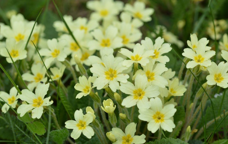
The right-hand photo shows the section at the equator and the medium thickness of the exine. The left-hand photo shows the pitted textured surface and the round pore apertures. The aperture pores look like little craters on the surface.
Spring Pollen Microscopy Summary
This weekend there was an abundance of spring forage. I was keen to take a closer look at the forage that the bees were working and bringing back to the hive, by doing some field photography and pollen microscopy.
It was evident that most of the pollen coming back to the hive was oil seed rape and dandelion. Smaller quantities of other pollen was being brought back to the hive, and that these included black thorn.
It was fascinating to see the different shapes and colours of the pollens. Rex Sawyer’s Pollen Identification for Beekeepers was also a great resource for understanding and classifying pollen.
If you have found this interesting or helpful, please subscribe to my blog, comment on the post, or like/share on Facebook.
Hi ,
Just a quick question , how are you finding your microscope set up from Apex ? We are just about to buy one , they seem very suited to purpose with the mini grabber and your photos look great.
Hi Anthony
I found the apex researcher really good for the BBKA microscopy preparation and exams. It is simple to set up and adjust and the optics are good. I found the eye cam plus really simple to use to and gave some good results. The key to great microscopy photos at the magnification you need for pollen work is layering software, which allows you to “stitch” together photos of the same object, to overcome the issue that the object is bigger than the depth of focus of the lens. I use Zerene Stacker but also looked a Helicon.
I have found the team at Brunel microscopes really helpful when I have rand them with questions. (Apex is a part of their business and you get the same person if you ring the Apex or Brunel number!)
Good luck with your journey into microscopy, it is fascinating!
Regards Ian